Comprehensive. Convenient. Compassionate.
A Division of Pomona Valley Hospital Medical Center
When a doctor needs to look inside the body to diagnose an injury or illness, imaging tests are often the next step. Radiology uses advanced technology to create detailed pictures of organs, bones, and tissues that cannot be seen during a physical exam.
These images help healthcare providers identify medical conditions, monitor disease progression, and evaluate how well treatments are working.
Radiology exams are commonly used to investigate symptoms such as pain, swelling, injuries, or unexplained changes in health.
Understanding the different types of medical imaging can help patients feel more comfortable when their doctor recommends a radiology exam.
Radiology includes several imaging technologies, each designed to provide different types of information about the body.
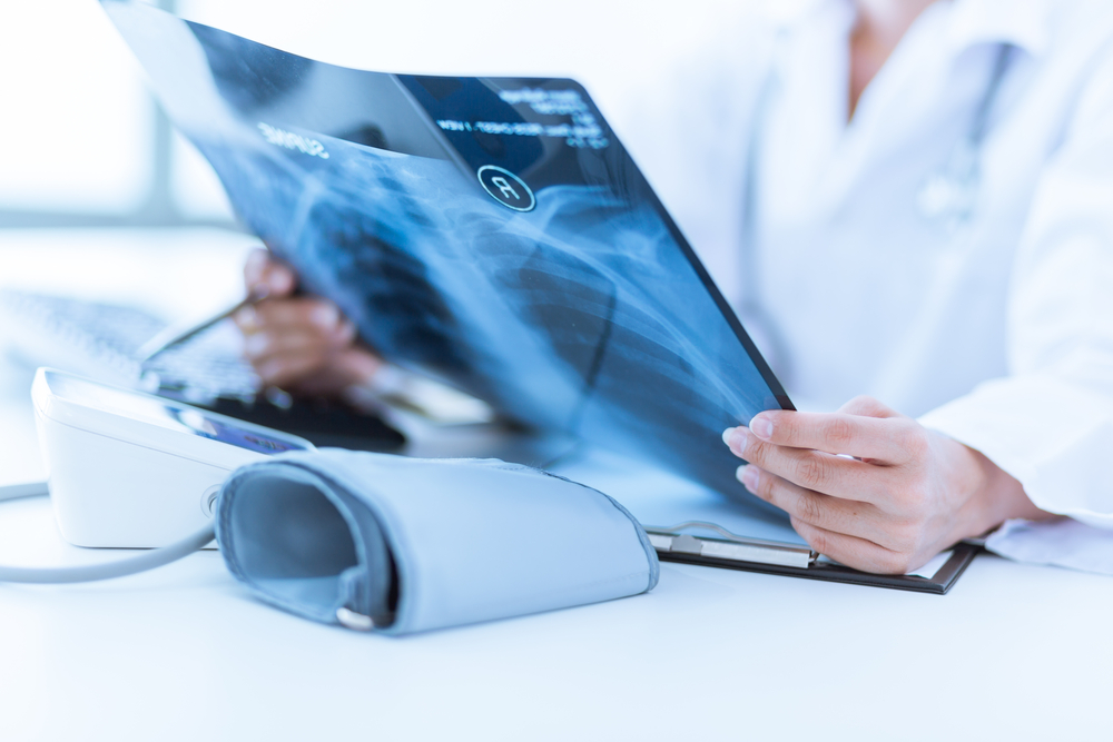
X-rays are one of the most widely used forms of medical imaging. This technology uses a small amount of radiation to create images of bones and certain internal structures.
Doctors commonly use X-rays to evaluate fractures, joint problems, lung conditions, and infections such as pneumonia.
Computed tomography (CT) scans combine multiple X-ray images to create detailed cross-sectional pictures of the body. These images allow physicians to see bones, organs, and soft tissues with greater clarity.
CT scans are often used when doctors need a more detailed view of internal structures, such as when evaluating injuries, infections, or possible tumors.
Ultrasound imaging uses sound waves to produce pictures of organs and tissues inside the body. Because ultrasound does not use radiation, it is commonly used during pregnancy to monitor the development of an unborn child.
Doctors also use ultrasound to evaluate blood flow, abdominal organs, and soft tissue structures.
MRI uses a powerful magnetic field and radio waves to produce detailed images of the body’s internal structures. This imaging technique is particularly useful for examining soft tissues such as the brain, muscles, joints, and spinal cord.
MRI scans are often used to evaluate neurological conditions, joint injuries, and certain internal diseases.
Mammography is a specialized form of imaging that uses low-dose X-rays to examine breast tissue. Screening mammograms help detect early changes in breast tissue that may indicate breast cancer, often before symptoms appear.
Early detection through screening plays an important role in improving treatment outcomes.
Nuclear medicine imaging uses small amounts of radioactive material, called radiotracers, to evaluate how organs and tissues function. These tracers travel through the body and are detected by special cameras that create images for physicians to analyze.
This type of imaging may be used to detect certain diseases at early stages or to evaluate how organs are functioning.
Radiology plays an essential role in modern healthcare because it allows doctors to diagnose conditions without surgery. Imaging tests help providers:
Because each imaging method offers unique insights, doctors choose the type of test that best fits a patient’s symptoms and medical history.
Medical imaging continues to advance, helping physicians detect conditions earlier and better understand what is happening inside the body. These technologies play an important role in diagnosis, screening, and monitoring many health conditions.
To explore how different imaging technologies support diagnosis and treatment, you can learn more about radiology services and how they are used across many areas of healthcare.